Specifications
 How the system operates
Future design options
 Blood / plasma mass densitometry
Standard inflight experimental protocol
 The "Chibis" LBNP device
 How the system operates
 Blood / plasma mass densitometry
 The "Chibis" LBNP device
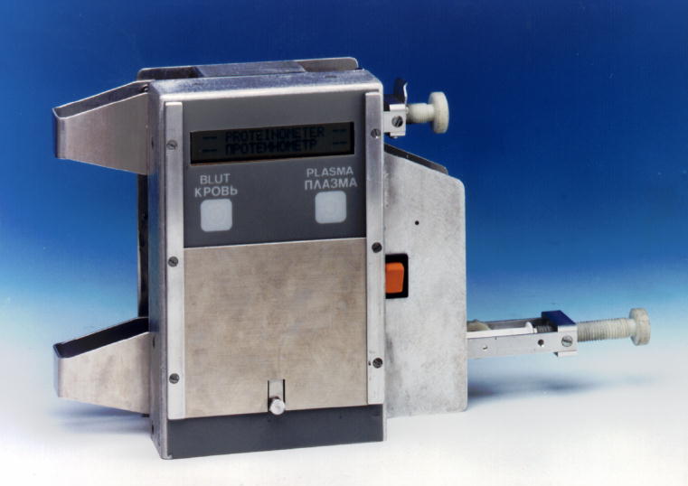
The ”Proteinometer” was especially designed for use in space and allows for sound travelling time (±1.0 µs) and temperature (±0.03 K) measurements on blood and plasma samples under the following environmental conditions:
The device offers two independent ports with different adapter mechanisms for blood and plasma measurements,
and consists basically of a whole blood measuring cell, a plasma measuring cell, the electronics unit, and an LCD display. Two push-buttons are provided for menu-directed operation. A sliding lid protects the buttons from damage during transport and stowage (”closed” position) or accidental activation during data retrieval (”medium” position).
The device employs a pair of piezoelectric transducers mounted to opposing positions in the wall of the cylindric measuring chamber, perpendicular to the length axis. Short acoustic pulses with 3 MHz center frequency are repeatedly transmitted through the sample and the propagation time is determined. The period of n cycles of a tunable voltage controlled oscillator (VCO) is adjusted to the referring propagation time the value of which is converted into a certain number of VCO cycles. A delay line is employed so that reflected and bypassed acoustic signals remain below a threshold value. The 2P-time of the VCO clock is mesaured by a cristal time base. The time measuring error in the electronic circuit is ~10 ns, the statistical error is in the order of 1%, ie ~100 ps.
A separate main switch activates the unit. The batteries are housed in a casing which fit in the Proteinometer in two positions, one for operation and one where no electrical contact is given, to prevent accidental power loss during transport or stowage.
Blood / plasma introduction is assisted by a syringe / couvette docking receptacle which comes with a lever guide designed for easy and leak-proof sample transposition. When not in use, the levers are folded in a protective transport position. The measuring cells are provided with a double piston-spring system designed for deadspace free (and therefore air-bubble free) operation and automatic sample removal / retrieval after measurement.
Sound velocity (m/s) in blood and its constituents is, like mass density (5), a linear function of protein concentration (1,2,7). From changes in blood and plasma, with hematocrit as a weighting factor, we computed sound velocity of the fluid (”filtrate”) which is exchanged between the flowing blood (intravascular compartment) and tissue / lymphatic spaces (extravascular compartment). Using this value, the volume of shifted fluid can be computed as % blood/plasma volume.
Typical propagation times in blood / plasma samples are about 10 µs, the accuracy therefore can be estimated as 10-5 (3). From the overall (measured) propagation time (Pm) which is the sum of a delay time T and the propagation time in the sample Ps, sound velocity SV is computed for a given propagation path L:
SV = L / (Ps - T)
Validation of measured SV data: [T] was derived by measuring Pm in samples (usually double destilled water) of known SV at certain temperatures. A control set was provided for routine reliability / precision checks using double destilled water and a control nomogram (3,7).
After preparation of the equipment (LBNP device, blood collection kit, centrifuge, SPVT device, plasma container, freezer), the test person entered the onboard LBNP (”Chibis”) device. Under 1-g conditions, the subject lay in a supine position during the whole experiment, allowing for establishment of supine steady-state conditions 60 minutes pre-LBNP. A venous indwelling catheter was inserted into the left antecubital vein. 3 minutes after starting the LBNP profile (-15/-30/-35mmHg for 15/15/10 minutes), the first blood sample was drawn. 2 minutes after termination of LBNP, the second blood sample was taken. This corresponds to previous ground-based findings of largest differences in plasma total protein and hemoglobin / hematocrit values (4). Whole blood SV was directly measured from a 2-ml sample drawn in a syringe which contained 500 IU heparin for anticoagulation. The other 2 samples (10 ml each) were double-centrifuged for plasma preparation and analysis on earth as described previously (6).
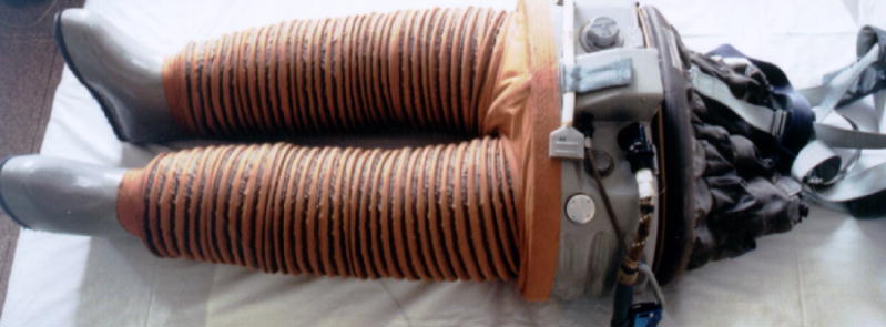
To create subatmorspheric pressure around the lower half of the body (lower body "negative" pressure = LBNP), the onboard "Chibis" device was employed for experimental purposes ("Bodyfluids / Interstitium"). LBNP is used inflight for countermeasure purposes (particularly before landing). LBNP creates a cardiovascular situation similar to upright standing on Earth, because blood is diverted from the central intrathoracic to the lower leg venous compartments. Thereby cardiac preload is reduced, just as with orthostasis (passive upright positioning) under 1-G conditions. As standing on Earth, application of LBNP in space can contribute to a cardiovascular training effect, which should ameliorate orthostatic stability upon landing, and reduce the risk of (pre)syncopy and loss of consciousness.

The Proteinometer was initially built for a strict single ("one-way") use within the frame of the Austromir project. As it turned out after F. Viehböck's return from his 9-day mission in 1991, cooperation between Russian (IMBP) and Austrian experts in the area of space life sciences was to be continued. But some of the AUSTROMIR hardware was not designed for long-term use, particularly the Proteinometer with its high-precision measuring chambers which in that case should have been meticulously cleaned after every single use.
It was decided to use the Proteinometer anyway, and a cleaning kit was designed and sent onto the MIR station. However, problems occurred which were due to the fact that the Proteinometer was not meant for continuous use. If the experiment planning would have allowed for the option of continuation of the scientific program, a different device would have been designed and constructed. One good option would be a high-precision mass density mesuring apparatus, employing the so-called mechanical oscillator technique; it has been very successfully used for determination of protein concentration changes in blood and plasma samples.
Mass density D is defined as mass per unit volume. Its determination with the mechanical oscillator technique (MOT) is based on the mass spring principle and high-resolution oscillation-time determinations. The actual resonant frequency ( f ) on an oscillating U-shaped glass tube with a certain filling volume is determined according to
f = 1/2 Pi.c [sqrt] ( Mo + D.V )
where c is the constant of elasticity for the oscillating tube, Mo is the inertial mass of the empty tube, and V is the volume of the sample contained in the tube.
The tube, with a piece of metal attached on its tip, is brought to bending-type oscillations perpendicular to the plane of the U by sinusoidal electromagnetic stimulation provided by the density measuring apparatus (DMA). The tube is fixed on two points which can be looked upon as the axis for the bending movements. Consequently, any sample must fill the tube within the two fixation points homogenously to give a reliable density reading.
A density measuring apparatus (DMA)-602 M (Paar KG, Austria) needs not more than a 0.1 ml sample volume within the measuring cell.
Selected publications